The number of people developing skin cancer over the course of their lifetime has risen sharply in the past 20 to 30 years. In Germany alone, it is estimated that there are over 280,000 new cases of skin cancer each year. Skin cancer mainly develops when the skin is exposed to excessive ultraviolet radiation — whether from natural sunlight or tanning beds. The face is most commonly affected by non-melanoma skin cancer (basal cell carcinoma), while melanoma, the more aggressive form, can occur anywhere on the body.
In addition to diagnosis and determining whether a tumour is benign or malignant, the professional treatment of skin lesions is always the primary focus of our care.
An increasing number of patients value the aesthetic outcome of tumour removal, whether benign or malignant. As specialists in plastic and facial surgery, we are committed not only to treating the condition but also to achieving an aesthetically pleasing result. When necessary, we use ultra-fine radiofrequency scalpels and precision suturing techniques to ensure the best possible outcome.
All tissue samples are processed in cooperation with a certified pathology institute linked to the hospital. Thanks to a dedicated transport service, we typically receive results within 24 hours, allowing for rapid diagnosis and treatment planning.
Depending on the extent of the procedure and the patient's wishes, surgery is usually performed under local anaesthesia but can also be carried out under sedation or general anaesthesia.
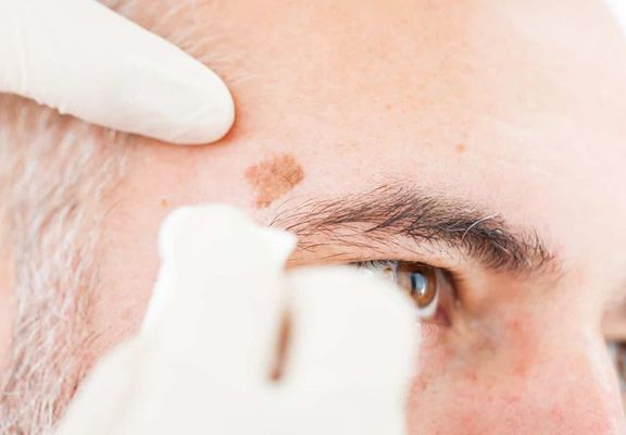
How can I tell what type of skin lesion I have?
It is often difficult to determine clinically whether a skin lesion is benign or malignant. For this reason, if any suspicious change is detected, we recommend taking a biopsy for histological (microscopic) examination in a pathology laboratory. Using specialised embedding and staining techniques, the pathologist can identify exactly what type of skin change is present.
How is surgical treatment of skin tumours performed?
This depends on whether it can already be clinically assessed if the skin lesion is benign or malignant. In cases of clearly benign skin tumours, removal and aesthetic reconstruction of the affected area can often be completed in a single appointment.
If the finding is potentially malignant or semi-malignant (such as a basal cell carcinoma), we recommend removing the lesion and leaving the wound open initially. After the procedure, the area is covered with sterile gauze and discreet, skin-coloured dressings until the pathology results are available. Once the results are received, we determine whether any remaining tissue needs to be removed or if the wound can be closed.
What types of benign skin lesions exist?
Tumours are often automatically associated with malignancy, but there are many skin lesions that are benign — medically non-cancerous. When such changes occur on the face, they can usually be removed safely and with aesthetically pleasing results. Examples of benign skin tumours include:
- Seborrhoeic keratoses (age warts)
- Birthmarks (haemangiomas or port-wine stains)
- Pyogenic granulomas
- Milia
- Moles (naevi)
- Haemangiomas
- Fibromas
- Granuloma annulare
- Lipomas
- Sebaceous cysts (atheromas)
- Sebaceous naevi
What types of malignant skin lesions exist?
Malignant or semi-malignant skin tumours occur when skin cells begin to grow abnormally. There are two main categories, each with several subtypes:
- Non-melanoma skin cancer (white skin cancer)
- Basal cell carcinoma (basalioma)
- Squamous cell carcinoma (spinalioma)
- Melanoma (black skin cancer)
What is white or non-melanoma skin cancer?
The two main forms of non-melanoma skin cancer are basal cell carcinoma (basalioma) and squamous cell carcinoma. Basal cell carcinomas are sometimes described as "semi-malignant" because they grow slowly and very rarely spread (metastasise) to other organs. When detected early and completely removed, the chances of full recovery are excellent — around 95%.
Basal cell carcinomas usually appear as flesh-coloured nodules on areas of skin frequently exposed to sunlight, such as the face.
Squamous cell carcinomas, on the other hand, are malignant skin tumours that often develop from actinic keratoses — benign but precancerous rough, reddish skin patches caused by long-term UV exposure. These also frequently occur on the face. If diagnosed and removed early, this form of skin cancer can usually be completely cured. Metastasis is rare.
What are the symptoms of non-melanoma skin cancer?
An actinic keratosis typically appears as a reddish-yellow, scaly patch of skin that may bleed slightly when scratched or picked. The surrounding skin often shows mild redness due to inflammation.
In advanced stages, squamous cell carcinomas become thicker and take on a whitish appearance. They may spread gradually and develop into wart-like, irregular skin growths. When touched, they often feel rough — similar to coarse sandpaper. Attempts to remove the hardened skin may cause bleeding.
Basal cell carcinomas usually begin as small, whitish-grey nodules only a few millimetres in size. These bumps may have a pearly sheen, and small blood vessels can often be seen on the surface. If the lesion is scratched or irritated, it may form a thin, bloody crust. Over time, the centre of the nodule may sink in, forming a small crater with a raised, rolled edge where tiny blood vessels are visible. The tumour gradually grows deeper into the surrounding tissue.
What is malignant (black) skin cancer?
The term "black skin cancer" refers to malignant melanoma, a type of skin cancer that develops in the melanocytes — the pigment-producing cells responsible for skin colour (melanin). Malignant melanomas may develop from existing moles (naevi) or from normal, previously unaffected skin. They often appear as dark, irregularly shaped patches or lesions on the skin. The tumour cells typically begin to grow in the lowest layer of the epidermis (the outer skin) known as the stratum basale. Malignant melanoma is particularly dangerous because it can metastasise — spreading to other organs if not detected and treated early. Early detection through regular skin examinations and careful observation of any skin changes is essential to ensure the best possible treatment outcomes.
What are the symptoms of malignant melanoma?
To distinguish malignant melanoma from other skin changes, dermatologists use the ABCDE rule as a practical guide. The following features may indicate a possible melanoma:
- A for Asymmetry: Melanomas usually grow unevenly. A mole with an asymmetric shape may be a warning sign.
- B for Border: Melanomas often have irregular, jagged, or blurred edges, with no clear transition between the affected and healthy skin.
- C for Colour: Malignant melanomas tend to have uneven colouring, ranging from reddish-brown and black to grey or even whitish tones.
- D for Diameter: Any mole larger than five millimetres in diameter should be checked by a dermatologist. Melanomas tend to grow progressively over time.
- E for Elevation: A melanoma may be raised above the surrounding skin surface. Dome-shaped or elevated pigmented lesions should always be considered suspicious.


